
The Art of Science On Display Under the Microscope
Beautiful images come to life during the detailed scientific process. Meet one scientist who captures the art.
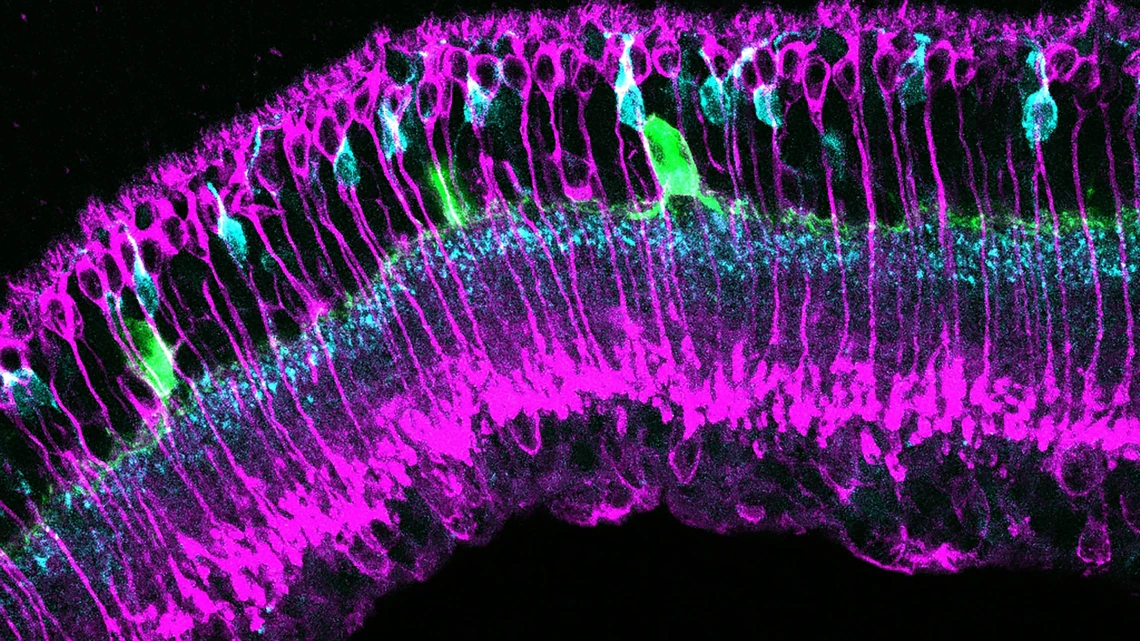
This image shows different types of retina cells labeled in green, magenta and cyan. Scientists are studying images like this from diabetic and control retinas to understand the cellular changes that occur in the early stages of diabetes, before vision loss occurs.
In addition to advancing critical research in medicine and science, one of the unintended outcomes of basic science is the beautiful imagery that’s only viewable under a high-powered lab microscope.
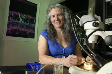
Researcher Andrea Wellington uses a dissecting microscope in a lab on main campus. An image captured while researching diabetes hangs in the background.
“Science is happening that nobody ever really sees. I mean, you might see it in the publication, but your typical non-scientist isn't going to have any idea the retina could be so pretty.”
Andrea Wellington is an assistant research scientist in the Eggers Lab in the Department of Physiology at the College of Medicine – Tucson. She also moonlights as an art hobbyist who photographs the biological art she sees under a confocal microscope.
Wellington studies diabetic retinopathy in mice to find targets for medications to help people with diabetes maintain their eyesight. While staining cells and observing the retina for changes due to the treatments, she is always struck by the colorful images staring back at her.
“There will be times when I'm sitting in the lab looking at what I'm imaging, and I think ‘Wow, this looks really cool.’”
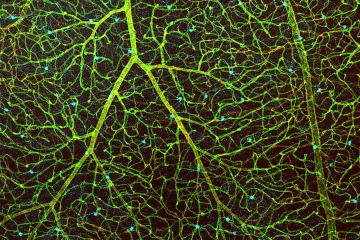
This image shows the blood vessels in a retina in green, the cells that make up the blood-retina barrier in red, and dopamine-producing amacrine cells in cyan.
Wellington first makes sure to capture all the images she needs for research purposes.
“When I'm done, I'll go take a quick look at the optic nerve head and see if there’s anything exciting. I'll just take a couple of minutes to save the images I thought were interesting.”
Six years ago, she started participating in Symbiosis, an annual biological art exhibition hosted by the UArizona Neuroscience and Cognitive Science Ambassadors. She has displayed some of her images at Symbiosis each year since then. One of her most recently exhibited images was printed on canvas as a prize for winning the Vector Labs’ photo contest.
Since then, she has submitted numerous other images from under the microscope to the Microscopy Society of America and to the Nikon Photo Contest.
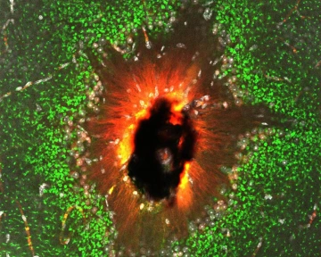
Image titled “The Eye of Sauron” captures the back of the retina.
One of Wellington’s recent images is a picture of where the optic nerve head comes out of the back of the retina, titled “The Eye of Sauron.” “People who do confocal microscopy realize that so many images are just gorgeous, and not everybody chooses to take the time to portray it as art,” said Wellington.
Today, evidence of her art hobby can be found all around her home office, including prints hanging on the wall, on coasters, and on coffee mugs, blankets and mouse pads.
Wellington said she often gives those items as gifts because they’re so unique. She gets positive reactions when sharing them on social media, even from people who aren’t familiar with the science.
“There’s so much science that's happening, and non-scientists don’t get to see it,” said Wellington. “Part of my motivation it is just the desire to take the time to share these beautiful images with the world.”
Photo Gallery: Scientific Research Reveals Microscopic Beauty