
Have 3-D printed brain; will travel
College of Medicine – Tucson team is creating its own brain models to help medical students better understand ‘normal’ anatomy.
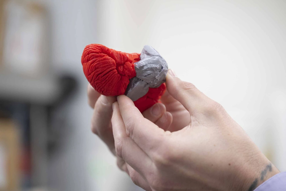
Creating 3D printed brain models helps students connect the virtual anatomy they see in a book or on screen with real human brain differences.
Photo by Noelle Haro-Gomez, U of A Health Sciences Office of Communications
Figuring out how the brain works is a complicated business. On the one hand, the brain controls the body’s autonomous systems, like our heart, lungs and digestive system. On the other, it is the home of thoughts, emotions and memories, those ephemeral sentiments that make us human.
But when there is something awry in the brain, it’s not just a simple task to create an image or cut it open to get a better look at what is happening. This is especially true when teaching medical students, who need to be able to see the inner parts of human anatomy without losing critical context that the outer layers provide.
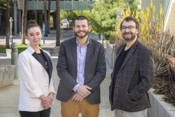
Haley O’Brian, PhD, Paul Gignac, PhD, and Micah Etter, MD, are the brains behind the 3D printing program for students in the U of A College of Medicine – Tucson.
Photo by Noelle Haro-Gomez, U of A Health Sciences Office of Communications
“Dissection is the gold standard, but it necessarily destroys part of the organism we are trying to understand,” said Paul Gignac, PhD, an associate professor in cellular and molecular medicine at the University of Arizona College of Medicine – Tucson, and the director of physician assistant anatomy at the College of Health Sciences. “We routinely turn to contrast-enhanced computed tomography, where we can see soft tissues or magnetic resonance imaging of those parts that we want to better understand. That gives us ‘slices’ of what’s being examined.”
Which is great, Gignac said, but when students need to connect the virtual anatomy with real anatomy, 3-D printing can come to the rescue.
With a 3-D printed body part, students can compare the radiology study from the CT or MRI and then a physical object to allow for a better connection between the two. This makes the concept more “sticky” for the health care student when reviewing case studies or learning how to diagnose patients.
Making “normal” anatomy the norm
“One of the topics that is really important to us to cover with our health care students is that there’s a pretty wide range of what’s normal,” Gignac said. “They need to be able to appreciate normal anatomy and differentiate it from abnormal or unhealthy anatomy. Traditional models usually give you an idealized form of what’s normal or healthy anatomy. But when we use anonymized patient studies, we can say this patient had a tumor or an aneurysm, and we can then 3-D print that structure, and students can hold it in their hands, which would be otherwise impossible to do. They can appreciate what an abnormal vascular system looks like, for example, and how it’s distributed, not just an artistic rendering of that vascular system. So, it connects the normal and abnormal anatomy more directly to what they will see in patient imaging studies, and it gives students a more intuitive sense of how to differentiate those two kinds of presentations.”
Gignac is a professional anatomist and evolutionary biologist, and he and his colleagues are helping students “stick” the learning by using a 3-D printer to create models of human brains from digital MRIs. But why 3-D-print brains when resin models already exist?
Digitally reconstructing 3-D printable brains flips the anatomy lesson on its head. In the past, students would deconstruct the anatomy as they dissected a cadaver. Now, research students working in the lab reconstruct a body by rebuilding it from the radiology data manifested in the 3-D brain.
“We’re really excited to push that as a way to connect more anatomically detailed, patient-based data sets with the variation in anatomy that the students learn,” Gignac said. “We don’t only use the prints, but by associating QR codes of the virtual model that the print came from, we can fully annotate important structures in a virtual environment. The students can go from learning via a model and then testing themselves on the virtual version. They have an opportunity to work in multiple modalities and get a very rich kind of understanding of the features of interest.”
Lose a brain? Just print another
The 3-D printing of anatomy can be much more economical as well.
“It’s a dollar’s worth of biodegradable plastic,” said Micah Etter, MD, an assistant clinical professor in neurology at the College of Medicine – Tucson, and co-director of the nervous system block. “But these can be well received, and there’s a lot to be said, especially for neuroanatomy, where it’s very esoteric and you don’t have very many hands-on experiences with a dissected brain. First of all, you can’t really take a real brain home with you” to study, he added. But a 3-D printed brain with an aneurysm? Have backpack, will travel.
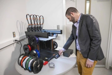
Paul Gignac, PhD, checks the progress of work set up on the 3D printer in his lab.
Photo by Noelle Haro-Gomez, U of A Health Sciences Office of Communications
“Part of understanding how it works is understanding how it moves,” Etter said. “And you can’t really understand how a picture in a book moves, or at least maybe not as readily as when I’m holding it in my hand and I’m spinning it clockwise and I’m seeing what’s happening. So, in that regard, I guess there are opportunities for different ways to appreciate the knowledge that is out there in a more readily accessible fashion for learners.”
Etter said he finds brain designs online — researchers openly share models they are working on — and augments them based on the needs of the lesson, whether it is adding an aneurysm or hair follicles for a model he is working on of the inner ear.
While it may be a less expensive learning tool, a quality print of a brain can take 10 to 12 hours, Etter said.
“The technology has improved quite dramatically over the past few years,” he said, “but it is also inherent to the kind of model you’re printing and the level of detail involved and the printing materials that are being used.”
The use of 3-D printing for anatomical models seemed a natural extension for Gignac.
“I’m an evolutionary biologist, and my research focuses on the evolution of the head,” he said. “I’m really interested in how the jaws and the brain dominate the way the head is organized. And in some groups of animals, as they become brainier, this tends to lead to smaller jaws. And in some groups of animals, they have big teeth and large jowls, and they tend to have smaller brains. And I probe this duality as part of my research program.”
A more accessible tool that travels better
Like many recent innovations in teaching, the COVID-19 pandemic accelerated the role of 3-D printing.
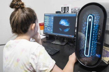
Emma Rose Buhman-Wiggs, a doctoral student in the Neuroscience Graduate Interdisciplinary Program, reviews a brain scan to use for a 3D print.
Photo by Noelle Haro-Gomez, U of A Health Sciences Office of Communications
“When students couldn’t come into the classroom, we had to really ramp up our approach to this,” Gignac said. “That was spearheaded by Haley O’Brien, PhD.” O’Brien is an associate professor of cellular and molecular medicine and associate head education in the College of Medicine – Tucson.
“The whole team turned to virtual models as ways for students to learn human anatomy. Now, students will come into the research lab, and they will learn about anatomy by taking these grayscale, volumetric CT and MRI scans, and they will create the digital models of the 3-D structures. They’re doing the inverse — instead of cutting away the structures to learn what’s underneath, they rebuild them from the radiology data, and they learn about the anatomy that way. We are embracing the way the tool allows you to kind of reconceptualize anatomy training.”
Gignac is working to expand access to 3-D models for the upcoming physician’s assistant programs in the College of Health Sciences.
“We want to build what’s called a bone box for groups of students,” he said. “This is a common feature of anatomy labs, where we have human skeletons that are full skeletons in boxes that the students will check out to study on the other side of the lab. They can’t leave the lab because the contents are Willed Body Program materials — human remains. When the lab is over, the students pack it all up, and they bring it back to where all the models and the bones are stored.
“Well, we‘d like to 3-D print whole skeletons. The students can then check one out and go home with it for the semester, and they can study on their own time. If a bone model is damaged, then we just 3-D print it again.”