
Researchers using $3.3M NCI grant to develop more accurate, comfortable breast cancer screening technology
College of Medicine – Tucson researchers are leading a team that will test noncompression CT technology, which has the potential to detect more breast cancers while increasing comfort and decreasing radiation exposure.
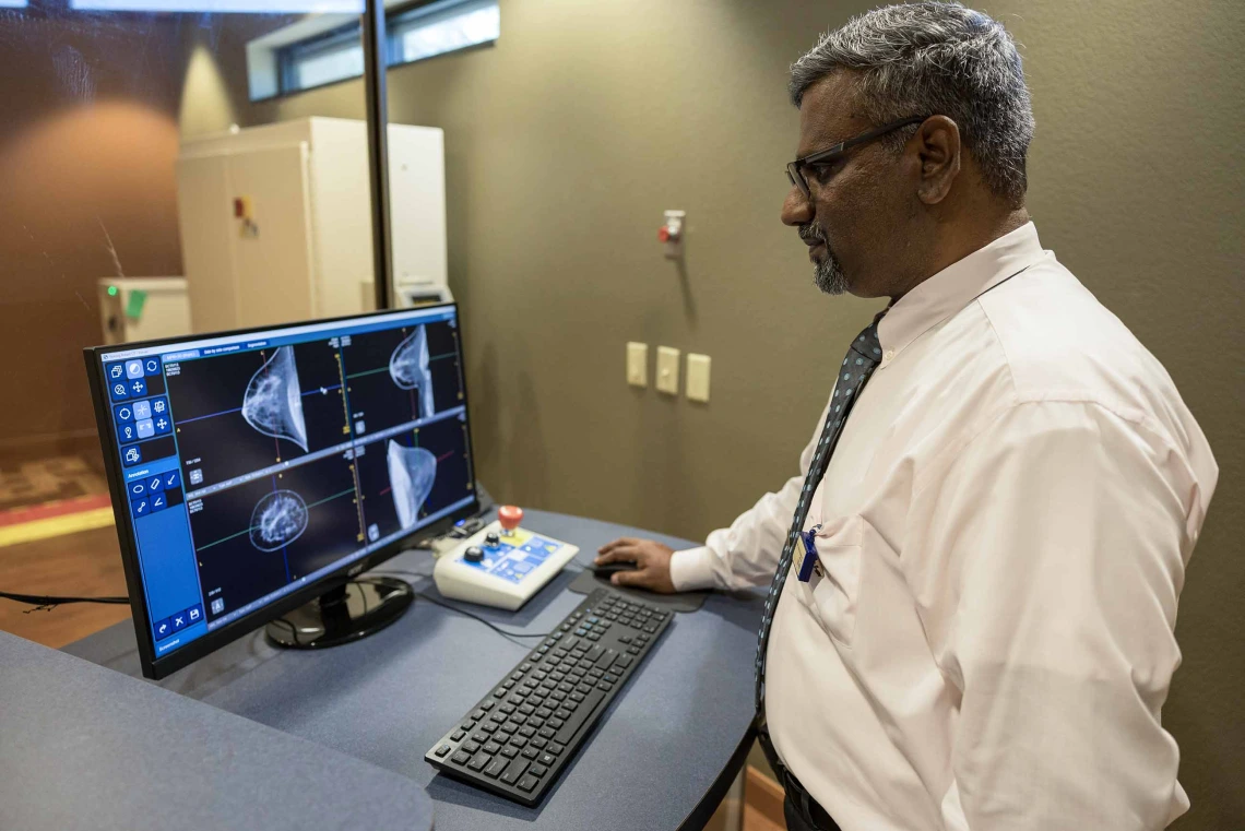
Srinivasan Vedantham, PhD, is leading a study that is testing CT imaging as an alternative to mammograms for breast cancer screening.
Photo by Quion Lowe and Lisa Dahm, U of A Cancer Center
Researchers at the University of Arizona Health Sciences are using a $3.3 million grant from the National Cancer Institute, a division of the National Institutes of Health, to continue testing a novel imaging method for breast cancer detection that they hope will one day provide a superior alternative to the mammogram.
The team’s advanced breast computed tomography, or CT, system does not require physical compression of the breast and eliminates tissue overlap. Instead, it uses detailed CT scans to create high-resolution 3-D images.
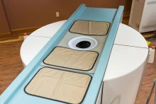
U of A Health Sciences researchers developed a prototype of a CT scanner that they hope will one day make breast cancer screenings more comfortable and effective than mammograms.
Photo by Quion Lowe, U of A Cancer Center
“With our technology, there is a hole on the table and the woman lies prone with the breast through the hole. The tube spins around 360 degrees,” said Srinivasan Vedantham, PhD, a professor in the U of A College of Medicine – Tucson’s Department of Medical Imaging and member of the U of A Cancer Center. “There is nothing in contact with the breast — no compression, nothing. You lie face down, and it takes 10 seconds to image each breast.”
Vedantham, who is leading the research team, said that in countries with national health services, such as the United Kingdom and Norway, a significant number of women of screening age who don’t receive regular screenings cite the pain of compression as the reason they avoid mammograms.
The team hopes this method could significantly enhance early cancer detection and diagnosis — and do a better job catching cancer in the half of women with dense breasts, which have more fibrous and glandular tissues that can obscure tumors.
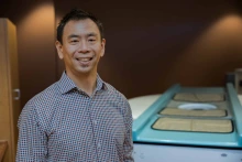
Alan Chiang, MD, PhD, an assistant professor of medical imaging and chief of the Division of Breast Imaging at the College of Medicine – Tucson, is a contributing researcher on the CT scanning project.
Photo by Quion Lowe, U of A Cancer Center
“The advantage of breast CT is we have a whole 3-D image. It would have better sensitivity in detecting breast cancer, particularly for women with dense breasts,” Vedantham said. “Our goal is to enhance breast cancer screening technology to improve early detection and outcomes for patients.”
Vedantham will use this grant to refine a noncompression CT scanner prototype that was tested with 92 women in collaboration with researchers from the U of A Cancer Center. This grant will focus on advanced image reconstruction techniques and recruiting 600 additional volunteers to help test the breast CT system, which will be compared with 3-D mammography, the current standard in breast cancer detection.
According to the NCI, breast cancer accounts for 15.5% of new cancer cases and 7% of cancer deaths in the United States. Approximately 13.1% of women will be diagnosed with breast cancer in their lifetime, making advancements in screening technology crucial. Though rare, some men develop breast cancer as well.
The applications of the team’s research extend beyond breast imaging and could someday be harnessed to improve radiation therapy, surgical planning and detailed imaging of other major organ systems.
Contributing researchers and collaborators include Alan Chiang, MD, PhD, assistant professor of medical imaging and chief of the Division of Breast Imaging; Sherry H. Chow, PhD, research professor of medicine and director of the Analytical Chemistry Shared Resource at the U of A Cancer Center; Denise Roe, DrPH, professor at the Mel and Enid Zuckerman College of Public Health and director of the Biostatistics Shared Resource at the U of A Cancer Center; and Hsin Wu Tseng, PhD, associate research scientist in the Department of Medical Imaging. The study is being conducted at the Banner – University Medicine Breast Imaging Center in Tucson and recruiting Banner patients who are being seen for routine mammograms.
This research is supported by the National Institutes of Health under award No. R01CA199044. Community members interested in participating in the study can learn more at clinicaltrials.gov.
Expert
Srinivasan Vedantham, PhD
Professor, Department of Medical Imaging, U of A College of Medicine – Tucson
Member, U of A Cancer Center
Contacts
Jennifer Fischahs
College of Medicine – Tucson
520-626-0011, jfischahs@radiology.arizona.edu
Anna Christensen
College of Medicine – Tucson
520-626-9964, achristensen@arizona.edu